Plant disease photography: what cameras can show and what a laboratory must confirm
How to read plant images, AI confidence scores and laboratory findings without confusing a visual change with a confirmed pathogen diagnosis.
Explore the guide
A camera records appearance. A model assigns an interpretation to an image. A diagnostic laboratory examines a defined sample using a particular method. These outputs can complement one another, but they are not interchangeable evidence.
Images document appearance
Colour, shape and visible texture are observations rather than a complete account of plant health.
Accuracy needs a setting
A result on a controlled image set does not establish performance in another facility.
A score is not confirmation
A model confidence score and a laboratory result describe different things.
What is actually in the image?
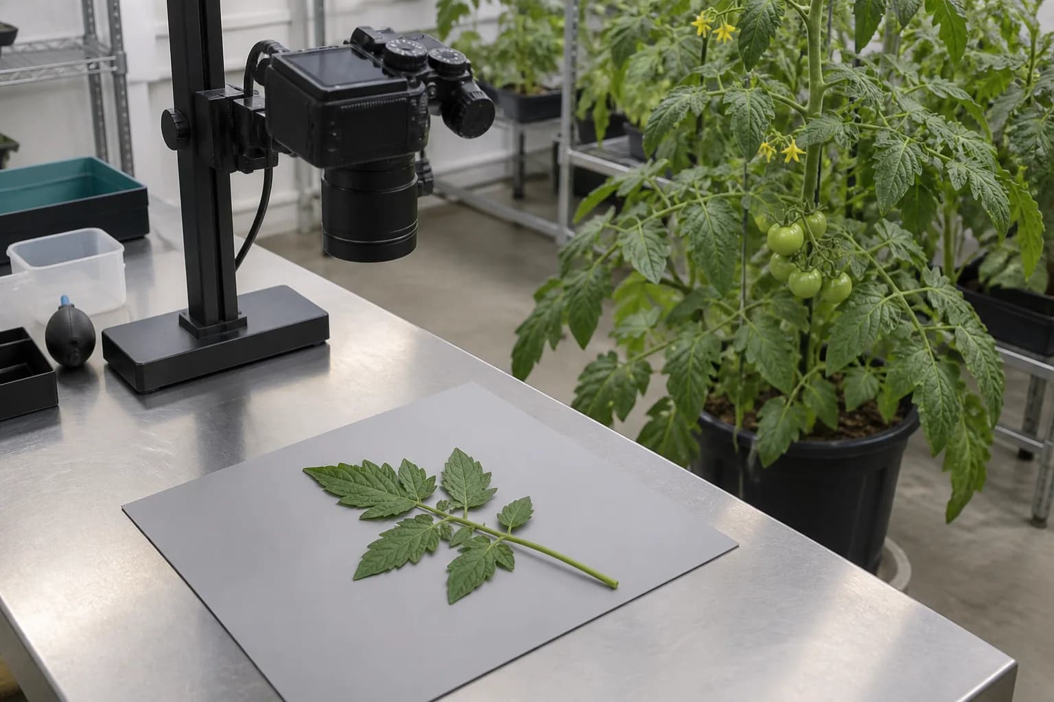
A leaf photograph contains the visible surface, lighting, viewpoint and background. It may show a change clearly while leaving the roots, internal tissues and earlier history outside the frame. University diagnostic guidance emphasises symptoms, their distribution and case history together. The professional value of an image is therefore not only its sharpness: it is also whether a reviewer can tell what it represents and what remains unseen. Diagnostic context.
The gap between a benchmark and a working environment
Mohanty and colleagues reported 99.35% accuracy on a held-out PlantVillage test set, but about 31% on two small external image sets. This 2016 result is not a current score for all plant AI, nor a cannabis validation. It demonstrates why changing image conditions can change performance. We discuss that experiment separately in our documented PlantVillage case. Original study.
Three outputs that should remain distinct
| Output | It can support | It cannot establish alone |
|---|---|---|
| Camera image | A visible change at a recorded time | Identity of every possible causal organism |
| Model suggestion | A category learned from training examples | Reliable performance outside its validation setting |
| Laboratory report | A method-specific finding in a submitted sample | A complete explanation of the whole facility |
What a laboratory result adds — and what it does not
A laboratory finding is tied to the submitted material, test target, method and reporting limits. It can add organism-specific evidence that an ordinary colour photograph cannot supply. It still needs interpretation in the context of the case. A targeted negative result is not a declaration that every possible pathogen is absent, and a positive finding does not automatically explain every visible symptom. University diagnostic perspective.
The interface should show the status of the claim
Hamfy’s proposed design principle is to keep an observation, an automated suggestion and a reviewed finding visibly distinct. A useful record can preserve the image, capture time, location identifier, model version and the separate external report. This is an information-design proposal, not a claim that Hamfy has validated disease recognition in cannabis. The pathogen overview explains why those distinctions matter.
Common questions
Does a 95% AI confidence score mean a diagnosis is 95% certain?
Not automatically. The meaning depends on the model, calibration, classes and deployment data. A displayed score should not be treated as a universal probability of disease.
Can we apply a published accuracy figure to Hamfy cameras?
No. Performance belongs to the tested model, dataset and evaluation. It must not be presented as Hamfy’s own result or transferred to a new crop and setting without evidence.
Is a photograph still useful when it cannot confirm a pathogen?
Yes. It can document visible changes and support specialist review. Its value is strongest when the image remains linked to the case history and the limits of the observation are clear.
Sources & research
Primary sources selected for this article. Reviewed by Hamfy on .
Evidence brief: camera observation versus diagnosisRead the research brief · scope, findings and limitations- Research source · 2016Using Deep Learning for Image-Based Plant Disease DetectionMohanty, Hughes & Salathé · Frontiers in Plant Science
Primary image-classification experiment; controlled and external evaluation are reported separately.
- Research source · 2024Field Notes talks crop disease diagnosis resourcesUniversity of Minnesota Extension
University plant-diagnostic guidance on symptoms, context and laboratory investigation.
Have a correction or additional project evidence? Contact the Hamfy team.
Connect observations with evidence.
Discuss a monitoring brief that keeps camera images, environmental records and verified findings in context.
Connected ideas
Cannabis plant pathogens: fungi, bacteria and the meaning of a diagnosis
Microbial contamination indoors: plant disease, environmental findings and product quality